Artificial intelligence (AI) is a relatively new and expanding field of research that can have beneficial applications in the world of medicine. Namely, AI is capable of analyzing data sets and learning how to diagnose cancerous tumours, proving it to be a beneficial tool for physicians. Positron emission topography (PET) is a form of nuclear imaging technology that provides a non-invasive way to analyze and diagnose malignant tumors (Sadaghiani, Rowe, and Sheikhbahaei 2021). PET imaging is based on labelling and identification of biomarkers, which then are measured through positron-emitting radionuclide tracers administered into the body (Griffeth 2005).
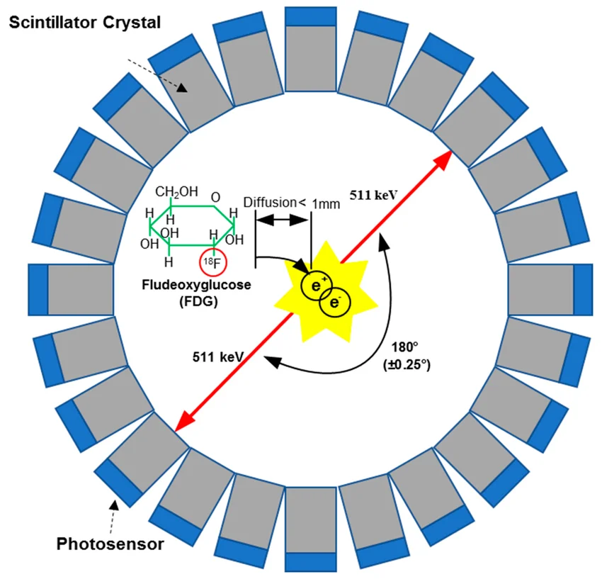
The most widely used radionuclide tracer for cancer detection is 18F-fludeoxyglucose (18F-FDG), as malignant cells have higher rates of aerobic glycolysis than in healthy tissues, requiring significantly more glucose to meet its energy needs. When 18F-FDG is administered into the body, it is phosphorylated to become FDG-6-phosphate in the cancer cell (Griffeth 2005). This causes it to become metabolically trapped, as the polar molecule cannot pass through the nonpolar cell membrane and exit the cell. This process causes accumulation of 18F-FDG in malignant cells, which allows the PET scan to locate the tumor.
Once administered intravenously, these radionuclide tracers undergo radioactive decay, with each decay event producing a positron, which travels less than a millimetre inside body tissues before undergoing an annihilation reaction with an electron. In this reaction, energy is released in the form of two 511-keV photons which travel in opposite directions (Griffeth 2005). These photons are then simultaneously detected by the PET scanner, forming an image that details tumor properties and allows for analysis of the tracer’s distribution. Images are produced through PET by surrounding the patient’s body with rings of specialized scintillator crystals and photosensors which detect the photons produced (Griffeth 2005), as shown in Figure 1.
PET scans, while being effective at cancer detection and analysis, are not 100% effective at proper diagnoses. The images must be interpreted by doctors, a process which is inefficient and can differ amongst doctors with different experience or knowledge, resulting in struggles in proper tumour diagnosis or prognosis (Huang et al. 2022). Additionally, other tissues can also show an increase in 18F-FDG activity, as infectious or inflamed tissues as well as changes to tissues post-surgery or chemotherapy can accumulate glucose and generate a false-positive scan result(Almuhaideb, Papathanasiou, and Bomanji 2011). This is observed often in both end-of-treatment and interim PET scans (Adams and Kwee 2016), as shown in Figure 2, and can be detrimental when combined with the potential for false negative scans due to human error from doctor analysis.

This issue can be remedied through data collection of previous PET scan results, which can be implemented into AI software, improving PET scan diagnosis accuracy. This is done through data learning (DL), a form of AI learning involving a set of methods that allows a machine to be fed raw data (Sadaghiani, Rowe, and Sheikhbahaei 2021). The AI then learns patterns based on these data sets through a convolutional neural network (CNN), a form of DL inspired by a neural system, where AI detects the necessary classifications that determine whether or not a tumor is malignant (Sadaghiani, Rowe, and Sheikhbahaei 2021). The application of DL in conjunction with PET scans has already proven to be highly effective in detection and assessment of survival and prognosis of cancer (Yuan et al. 2024), proving it to be a crucial tool in clinical decision-making.
Overall, the utilization of AI to assist in cancer diagnosis and prognosis proves to be an asset for physicians and a significant advancement in our capabilities of diagnosing and treating cancer. Data collection and analysis is a key tool in the diagnosis of cancer through PET scans, which can be improved through AI applications. Through the use of DL, it is evident that proper collection of data sets and their integration into AI is a crucial technology, and in conjunction with the use of PET scans, has the capability to transform the future of oncology.
Bibliography
Adams, Hugo J.A., and Thomas C. Kwee. 2016. “Proportion of False-Positive Lesions at Interim and End-of-Treatment FDG-PET in Lymphoma as Determined by Histology: Systematic Review and Meta-Analysis.” European Journal of Radiology 85 (11): 1963–70. https://doi.org/10.1016/j.ejrad.2016.08.011.
Almuhaideb, Ahmad, Nikolaos Papathanasiou, and Jamshed Bomanji. 2011. “18F-FDG PET/CT Imaging in Oncology.” Annals of Saudi Medicine 31 (1): 3–13. https://doi.org/10.4103/0256-4947.75771.
Griffeth, Landis K. 2005. “Use of Pet/Ct Scanning in Cancer Patients: Technical and Practical Considerations.” Baylor University Medical Center Proceedings 18 (4): 321–30. https://doi.org/10.1080/08998280.2005.11928089.
Huang, Brian, John Sollee, Yong-Heng Luo, Ashwin Reddy, Zhusi Zhong, Jing Wu, Joseph Mammarappallil, et al. 2022. “Prediction of Lung Malignancy Progression and Survival with Machine Learning Based on Pre-Treatment FDG-PET/CT.” EBioMedicine 82 (August). https://doi.org/10.1016/j.ebiom.2022.104127.
Jiang, Wei, Yamn Chalich, and M. Jamal Deen. 2019. “Sensors for Positron Emission Tomography Applications.” Sensors 19 (22): 5019. https://doi.org/10.3390/s19225019.
Sadaghiani, Mohammad S., Steven P. Rowe, and Sara Sheikhbahaei. 2021. “Applications of Artificial Intelligence in Oncologic 18F-FDG PET/CT Imaging: A Systematic Review.” Annals of Translational Medicine 9 (9): 823–23. https://doi.org/10.21037/atm-20-6162.
Yuan, Lili, Lin An, Yandong Zhu, Chongling Duan, Weixiang Kong, Pei Jiang, and Qing-Qing Yu. 2024. “Machine Learning in Diagnosis and Prognosis of Lung Cancer by PET-CT.” Cancer Management and Research Volume 16 (April): 361–75. https://doi.org/10.2147/cmar.s451871.
Leave a Reply
You must be logged in to post a comment.