New, non-invasive methods of Early Endometriosis Detection through Vibrational Spectroscopy

Endometriosis is a chronic disease defined by growth of endometrial tissue outside the uterus, causing symptoms including but not limited to amplified pain throughout the menstrual cycle, pelvic discomfort, and infertility (Zondervan et al. 2020). The most effective way to diagnose endometriosis is through surgery, which is typically invasive, time consuming, and not often approved for younger patients (Humphries 2026). Late-stage endometriosis can be potentially diagnosed non-invasively through ultrasound, but early-stage endometriosis is not visible through imaging (Menakaya et al. 2026).

New strides have been taken in endometriosis diagnostic tools, thanks to patterns occurring in the chemical make-up of those with endometriosis. CA-125 (cancer antigen) and CA-19-9 are biomarkers used in detection of endometrial cancer. Upon study of ovarian tissue in endometriosis patients, there is a strong correlation between the CA-125 and CA-19-9 proteins in endometrial tissue and the presence of endometriosis in the body (Pant et al. 2023). Studies have shown that high levels of CA-125 and CA-19-9 combined in a patient yield about a 96% correlation to endometriosis progression in the body (Dai et al. 2015). That is, before endometrial lining grows to become late-stage endometriosis, identification of these biomarkers has potential in diagnosing and treating endometriosis before its symptoms become detrimental to the patient’s health.

Through the use of vibrational spectroscopy, biomarkers of endometriosis can be effectively identified, and patients can be diagnosed at faster rates than ever. Spectroscopy is a scientific study which identifies substances typically through their interactions with light waves. Vibrational spectroscopy is a non-invasive technique that measures the vibrational energy levels within a molecule through sound waves ( ScienceDirect Topics, n.d.).

In a study measuring vibrational spectra of a control group of healthy individuals against a group of people with diagnosed endometriosis, there was a significant difference in the wavelengths emitted between each group. Absorbances of biomarkers were measured in the study and plotted against each other in the graph below. (Olcha et al. 2026).
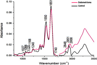
Figure 1-A graph plotting the absorbance against wavelength of biomarkers detected in the control group (black line) against the test group (pink line) (Olcha et al. 2026).

Through vibrational spectroscopy, promising advances in non-invasive diagnosis of endometriosis are exhibited. Moving away from invasive surgery as the primary way of diagnosing the disease will aid in controlling its impact as it grows over an individual’s course of menstruation. The average of diagnosis of endometriosis ranges from 5-12 years from the onset of initial symptoms due to the intrusive methods used in treatment of the disease (De Corte et al. 2025). With new developments in vibrational spectroscopy in the gynaecological field, individuals should hope to see earlier diagnosis periods of this chronic disease.

References

Dai, Xinyue, Chu Jin, Yan Hu, et al. 2015. “High CA-125 and CA19-9 Levels in Spontaneous Ruptured Ovarian Endometriomas.” Clinica Chimica Acta 450 (October): 362–65. https://doi.org/10.1016/j.cca.2015.09.019.

De Corte, Pauline, Moritz Klinghardt, Sophia von Stockum, and Klaas Heinemann. 2025. “Time to Diagnose Endometriosis: Current Status, Challenges and Regional Characteristics—A Systematic Literature Review.” Bjog 132 (2): 118–30. https://doi.org/10.1111/1471-0528.17973.

Humphries, Leigh Ann. 2026. “Endometriosis Surgery.” Obstetrics & Gynecology 147 (2): 169–73. https://doi.org/10.1097/AOG.0000000000006151.

Menakaya, Uche A., Kingsley N. Agholor, Dalia El Haieg, et al. 2026. “Endometriosis in African Women: Findings from a Cohort of Reproductive-Age Women Attending an Endometriosis Ultrasound Training Program.” International Journal of Gynecology & Obstetrics 173 (1): 545–47. https://doi.org/10.1002/ijgo.70606.

Olcha, Piotr, Wiesław Paja, Michał Kępski, et al. 2026. “Detection of Peritoneal, Ovarian, and Bowel Endometriosis Using FTIR Spectroscopy and Machine Learning.” Spectrochimica Acta Part A: Molecular and Biomolecular Spectroscopy 348 (March): 127242. https://doi.org/10.1016/j.saa.2025.127242.

Pant, Anuja, Kareena Moar, Taruna K. Arora, and Pawan Kumar Maurya. 2023. “Biomarkers of Endometriosis.” Clinica Chimica Acta 549 (September): 117563. https://doi.org/10.1016/j.cca.2023.117563.

“Vibrational Spectroscopy – an Overview | ScienceDirect Topics.” n.d. Accessed November 7, 2026. https://www-sciencedirect-com.libaccess.lib.mcmaster.ca/topics/medicine-and-dentistry/vibrational-spectroscopy.

Zondervan, Krina T., Christian M. Becker, and Stacey A. Missmer. 2020. “Endometriosis.” New England Journal of Medicine 382 (13): 1244–56. https://doi.org/10.1056/NEJMra1810764.

Comments

8 Responses to “New, non-invasive methods of Early Endometriosis Detection through Vibrational Spectroscopy”

  1. Joud Almadani Avatar
    Joud Almadani

    Hi iSci,

    Thanks for reading my post! I have wanted to write about endometriosis as an individual diagnosed with it, knowing that it does not get enough attention for how many people struggle with it daily. This new method of spectroscopy based diagnosis reminded me of our recent unit in chemistry on electrons in atoms and the photoelectric effect, as well as our research in RP 1 on spectroscopy to understand the chemistry on Titan! I hope you enjoyed reading.

    Joud 🙂

  2. Amelia Nolan Avatar
    Amelia Nolan

    Hey Joud!

    This is a great post. I did not know that our methods of spectroscopy were so early in development and were so complicated to diagnose. Here are a few quick suggestions to refine your draft:

    – Double-check your blog title capitalization; I’m not sure if “Early Endometriosis Detection” should be capitalized or not.
    – I noticed there is a minor space typo in your in-text ScienceDirect citation in P3.
    – You have a great figure! Make sure you reference it just before the image. (ie. “(see Figure 1)” could be placed P4S2). Also, ensure there is no period separating that last sentence and your reference in P4S2.
    – Additionally, your figure caption repeats some text from the blog. Consider elaborating slightly on what the absorbance differences between the control and test groups actually suggest (eg. higher and lower absorbance levels between groups at specific wavenumbers represent changes in the concentration and chemical structure of specific biomolecules).
    – As well, I would expand slightly more on statistics for the presence of biomarkers, such as what is considered “high levels” of CA-125 and CA-19-9, or what this is indicative of.

    You did a great job connecting spectroscopy from RP1 to endometriosis. Looking forward to reading your final draft!

    Amelia Nolan

  3. Jhaanvi Patel Avatar
    Jhaanvi Patel

    Hey Joud!

    I really enjoyed reading your post! I think you chose a really important topic, especially since endometriosis can be so difficult to diagnose and often goes unrecognized for years. Here are a few suggestions that I have:

    – In P2S2, I would clarify the distinction between endometrial cancer and endometriosis to differentiate between the conditions a bit more. The wording here makes it sound as though CA-125 and CA-19-9 are established diagnostic markers for endometrial cancer. I think stating that they are proteins that have been investigated as potential biomarkers for endometriosis would improve accuracy.

    – In P3S2, I noticed that vibrational spectroscopy is described as measuring molecular vibrations through sound waves. Since the study you discuss uses FTIR spectroscopy, I would clarify that this technique uses infrared light, which is absorbed at frequencies corresponding to molecular bond vibrations.

    – In P4S2, just a small punctuation adjustment: remember to remove the period after “graph below” and place it after the in-text citation instead.

    – Lastly, in the final paragraph, I would change “The average of diagnosis of endometriosis” to “The average time to diagnosis of endometriosis.” I think this makes the sentence flow a bit more naturally.

    Overall, amazing blog post! I love that you chose to focus on a condition that does not always receive the attention it deserves. Looking forward to seeing your final draft!

  4. Ayn Son Avatar
    Ayn Son

    Hi Joud!

    This is an amazing post! I never knew that methods such as spectroscopy could be used for diagnosis; you’ve definitely given me lots to think about. Here are a few suggestions to refine your draft:

    – P2: You mention that CA-125 is a cancer antigen, but you don’t explicitly state that CA-19-9 is one as well. Since CA-19-9 is not common knowledge, I suggest clarifying that both are cancer antigens. For example, you can phrase it as, “Cancer antigens CA-125 and CA-19-9 are….”

    – P3S3: I would double-check this citation, as citing ScienceDirect as an author is generally discouraged. Since ScienceDirect generates definitions from other texts (which I believe is where you got this citation), I suggest checking the source listed on the topics page and citing it instead.

    – P4: Here, I don’t believe the sentence “Absorbances of biomarkers…” is necessary, as you say a very similar thing in your figure caption. Instead, I suggest going into further detail about the study!

    – P4: You should mention your figure before it appears in the text.

    – P4: Finally, you have an extra period between your final word and your citation here.

    – P5S3: Here, you say “The average of diagnosis…” This is hard to follow; I would suggest saying, “The average time to diagnosis…”

    Overall, what a wonderful and informative post! I look forward to reading your final draft.

    Ayn 🙂

  5. Albesa Fazliu Avatar
    Albesa Fazliu

    Hi Joud!

    Extremely important topic! Advancements for endometriosis has not been treated with enough attention. I think this was a great topic to talk about, and I especially liked how you managed to connect aspects of chemistry from RP1 to it.

    I have some suggestions if you would like to use them:
    – P2S3: To make your sentence flow a bit better, you could change “Upon study of ovarian tissue in endometriosis patients” to “Studies of ovarian tissues from patients with endometriosis have shown..” so it flows slightly better.
    – I noticed after paragraph 3 that your reference has no date, this is a bit too meticulous but it’s a good habit to only use dated scientific sources for future writing.
    – For paragraph 4, if mentioned in your article regarding the study, I think it would be helpful to specify what samples were actually analyzed in the sample (for example, blood or tissue). Since your main argument focuses on the non-invasive diagnosis, this would make it clearer to the reader how the technique could eventually be used on patients.
    – You should implement your figure in text somewhere in the writing, potentially after P4S1 you can add: “there was a significant difference in the wavelengths emitted between each group (see figure 1)”.
    – In P5S3: You write, “The average of diagnosis of endometriosis ranges from 5-12 years…” I think you can refine this sentence a bit better, for example: ” The average time to diagnosis of endometriosis ranges from 5-12 years…”

    I can’t wait to read your final copy, best of luck editing!

    -Albesa Fazliu

  6. Farwa Rehan Avatar
    Farwa Rehan

    Hey Joud,

    Amazing post and a topic that is so important for so many women! I have a couple of suggestions to make your blog post even better:

    (1) Make your figure caption more descriptive. A figure should be able to stand-alone with its figure caption and still relay all the important information – this is especially important for graphs!
    (2) This is just a general note, but there should be no citations in the last paragraph of your blog post. No new information should be introduced.

    Once again, great job and great topic (I love women’s health)! I can’t wait to read the final copy.

    Happy editing!
    Farwa 🙂

  7. Nour Al-Sakka Avatar
    Nour Al-Sakka

    Hi Joud,
    I really enjoyed reading your blog post!
    Here’s some suggestions to consider while editing:

    – Make sure to centre your figure and refer to it in your paragraph before its displayed
    – Italicize your figure caption
    – I would double check the (Science Direct Topics, n.d) in-text, to make sure that it is correctly formatted

    Overall, great job. Looking forward to seeing the final product 🙂
    – Nour

  8. Sara Osman Avatar
    Sara Osman

    Hi Joud,

    This was a very interesting and informative blog post. I particularly liked how you highlighted a real-world medical challenge and introduced an emerging technology that could potentially improve early diagnosis. Here are a few suggestions:
    – The explanation of vibrational spectroscopy could be expanded slightly. You describe it as measuring molecular vibrations through sound waves, but could clarify how light interacts with these molecular vibrations to help identify specific biomarkers.
    – You mention a 96% correlation between CA-125 and CA-19-9 levels and endometriosis progression. It would be helpful to briefly clarify what this percentage represents and whether it refers to diagnostic accuracy, as correlation and diagnostic accuracy are not necessarily the same.
    – Your conclusion is promising, but could acknowledge that vibrational spectroscopy is still an emerging diagnostic technique and requires further research before it can replace existing diagnostic methods. This would provide a little more scientific context while maintaining your optimistic ending.

    Great blog post. Happy editing!
    Sara

Leave a Reply