The nude mouse was discovered spontaneously in 1966 by S.P. Flanagan from the Institute of Animal Genetics in Edinburgh, Scotland. The mouse was named “nude” after its phenotypically unique trait of hairlessness that arose in an albino strain named “nude” (Figure 1). These mice had increased postneonatal mortality, decreased reproductive success, and, in most mice, an inborn error of metabolism leading to the development of toxoplasmosis. It was later discovered in 1968 that these nude mice lacked a thymus, the immune organ that is in charge of T cell development (Mecklenburg et al. 2001). T cells are a group of lymphocytes that play a vital role in the adaptive immune system (Sauls et al. 2023). Because the nude mice have an underdeveloped thymus, they lack proper T-cell immunity. This explains their impaired defence against pathogenic organisms. These characteristics are due to a naturally occurring mutation in a gene called FOXN1. This gene regulates the development of certain tissues included in the hair follicles and thymus (Mecklenburg et al. 2001). Although this may seem like a large biological disadvantage, these characteristics have made the nude mouse extremely useful in scientific research. These animals have helped researchers investigate experiments and even potential medical treatments in fields such as oncology and immunology.
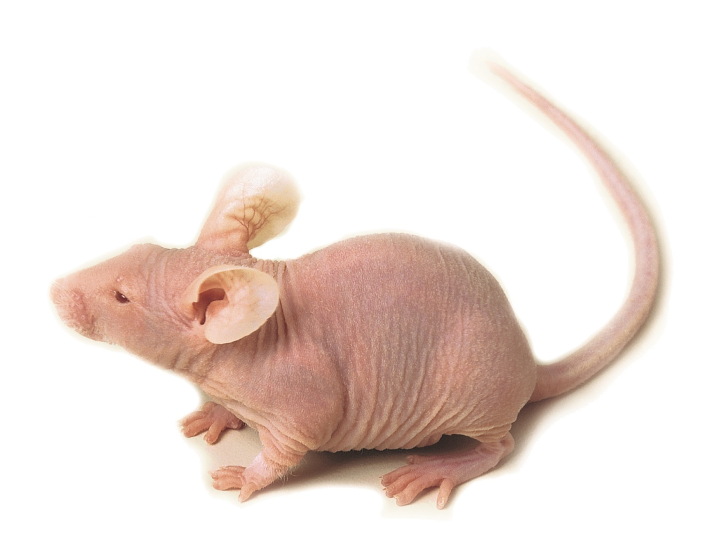
One of the most important applications for the nude mouse is in cancer research, especially in the use of xenografts. Xenografts are when tissue is transplanted from one organism into another organism (Zileli et al. 2017). In this context, tissue from cancer tumours can be transplanted into nude mice to run studies on the tumours. Since the mice have depleted T-cell immunity, they are less likely to reject the tumour tissue, as other animal models often would, making it an easier model to experiment on (Panaampon et al. 2021).
An early study run by Bellet et al. in 1979 tested whether nude mice could be used to predict how human melanomas respond to different cancer chemotherapeutic agents. In this study, they transplanted human melanoma cells into nude mice and exposed the mice to four different cancer drugs, two of which they knew had significant clinical activity and the other two were inactive. Their findings showed agreement with what was already established as an effective drug. The researchers suggested that the nude mice could be used as an effective preclinical screening tool for cancer treatments. However, the researchers had only tested 7 human melanoma cell lines, so further research was needed to determine the reliability of the model (Bellet et al. 1979).
Later studies applied this by using the same method while also supressing other immune cells. For example, a study done by Panaampom et al. in 2021 looked at the effectiveness of nude mouse xenograft models that lack Natural Killer (NK) cells, a type of cell that is important in the innate immune system. They created another kind of immunodeficient mouse called the nude-J mouse that were nude mouse containing a mutation the Jak3 gene, a gene that disrupts immune cell development. The findings showed that NK cells play an important role in rejecting tumour growth and that nude-J mice were optimal for the successful establishment of transplanted tumour cells(Panaampon et al. 2021).
Although these models are useful, there are some limitations that are present. The major limitation of nude mouse cancer models is that they do not reflect the behaviour of naturally occurring cancers in humans, so it is hard to compare what happens to the mice with humans. The nude mice have a deficiency in T cells; therefore, they do not mimic the response of a human immune system (Cekanova and Rathore 2014).
The nude mice show how an unusual genetic mutation can turn into a useful tool for scientific discovery. Even though it does not perfectly represent human physiology, it is still a useful tool in biomedical research that is widely used to this day.
References
AnLab. n.d. “Mutant Mice.” AnLab. Accessed November 20, 2026. https://www.anlab.eu/offer/laboratory-animals/laboratory-mice/mutant-mice/.
Bellet, Robert E., Victoria Danna, Michael J. Mastrangelo, and David Berd. 1979. “Evaluation of a ‘Nude’ Mouse-Human Tumor Panel as a Predictive Secondary Screen for Cancer Chemotherapeutic Agents.” JNCI: Journal of the National Cancer Institute 63 (5): 1185–88. https://doi.org/10.1093/jnci/63.5.1185.
Cekanova, Maria, and Kusum Rathore. 2014. “Animal Models and Therapeutic Molecular Targets of Cancer: Utility and Limitations.” Drug Design, Development and Therapy 8 (October): 1911–22. https://doi.org/10.2147/DDDT.S49584.
Mecklenburg, L., M. Nakamura, J. P. Sundberg, and R. Paus. 2001. “The Nude Mouse Skin Phenotype: The Role of Foxn1 in Hair Follicle Development and Cycling.” Experimental and Molecular Pathology 71 (2): 171–78. https://doi.org/10.1006/exmp.2001.2386.
Panaampon, Jutatip, Kenichi Sasamoto, Ryusho Kariya, and Seiji Okada. 2021. “Establishment of Nude Mice Lacking NK Cells and Their Application for Human Tumor Xenografts.” Asian Pacific Journal of Cancer Prevention : APJCP 22 (4): 1069–74. https://doi.org/10.31557/APJCP.2021.22.4.1069.
Sauls, Ryan S., Cassidy McCausland, and Bryce N. Taylor. 2023. “Histology, T-Cell Lymphocyte.” In StatPearls. StatPearls Publishing. http://www.ncbi.nlm.nih.gov/books/NBK535433/.
Zileli, Mehmet, Edward C. Benzel, and Gordon R. Bell. 2017. “166 – Bone Graft Harvesting.” In Benzel’s Spine Surgery, 2-Volume Set (Fourth Edition), edited by Michael P. Steinmetz and Edward C. Benzel. Elsevier. https://doi.org/10.1016/B978-0-323-40030-5.00166-0.
Leave a Reply
You must be logged in to post a comment.