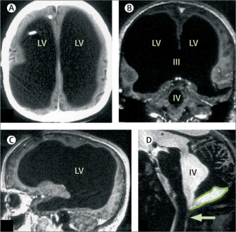
The 2007 report “Brain of a white-collar worker” by Dr. Lionel Feuillet, Henry Dufour, and Jean Pelletier, describes the case of a 44 year old with a history of childhood postnatal hydrocephalus. His CT and MRI scans revealed massive enlargement of his ventricles (Figure 1) (Feuillet et al. 2007). A few weeks after a ventriculoperitoneal shunt was inserted, the neurological examination displayed normal results. How was he able to live a normal life with so much of his brain ‘missing’?
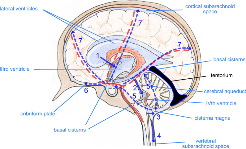
Hydrocephalus is a condition where the build up of cerebrospinal fluid (CSF) in the ventricles puts pressure on the brain (Mayo Clinic 2023). Too much fluid can lead to damage in brain tissue, and in severe cases the cerebral cortex is reduced to a ribbon of tissue (Mayo Clinic 2023; Ferris et al. 2019). Physiologically, the choroid plexus produces CSF within the lateral, third, and fourth ventricles (Figure 2) (Koleva and De Jesus 2023). CSF travels from the lateral ventricle to the third ventricle via the foramen of Monro. Then, CSF uses the cerebral aqueduct to flow to the fourth ventricle, from which it enters the basal cisterns. Part of it continues to flow around the spinal cord (Koleva and De Jesus 2023).
The CSF flow dynamics must be considered from the lens of fluid dynamics. Bernoulli’s principle explains that a fluid does not flow along a gradient of pressure or velocity, but along a gradient of energy, from a high (choroid plexus) to low (venous blood) energy region through the ventricles (Schmidt et al. 2016). Pressure and velocity are a part of the total energy. Ventricular expansion can occur after manipulation of intraventricular pulsatility and increased pressure pulse wave. This is because increase in “pulsatile energy” means ventricles have to somehow accommodate this change in total energy (Schmidt et al. 2016). This expansion of ventricles also means subsequent changes in volume. According to the Monro-Kellie doctrine, an increase in the total volume of CSF means a decrease in the volume of the brain and blood within the skull, since the total volume must remain constant (Koleva and De Jesus 2023). Without this, the intracranial pressure increases. This can cause brain damage and pressure induced atrophy (Koleva and De Jesus 2023).
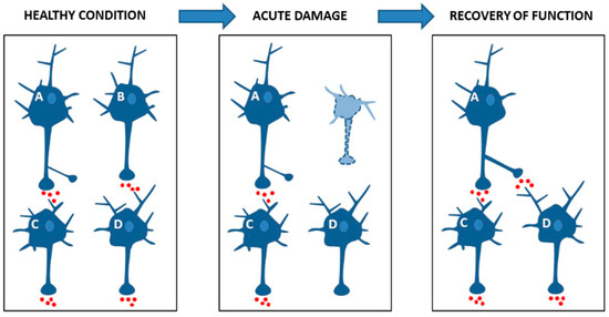
Neuroplasticity may potentially explain how regular functions can be retained, despite this increase in intracranial pressure and accompanied brain damage. It is the ability of the nervous system to reorganize its structure, functions, or connections after injury (Puderbaugh and Emmady 2023). One of the mechanisms of neuroplasticity is functional reorganization. When one area of the brain is damaged, another area of the brain is able to sustain this lost function (Figure 3). This is easier for children than adults when it comes to certain functions like speech (Puderbaugh and Emmady 2023). Consequently, early hydrocephalus may allow the brain to overtake lost functions, potentially explaining what may have occurred in the case of this 44 year old.
The case of hydrocephalus reported back in 2007 challenges our current ideas of brain injury. Understanding hydrocephalus through anatomy and physiology, the cause of damage through biophysics, and brain resilience through neuroplasticity, is important. This understanding is especially pertinent to future research on how the brain is able to retain function despite having extensive damage.
References
Ferris, C. F., X. Cai, J. Qiao, et al. 2019. “Life without a Brain: Neuroradiological and Behavioral Evidence of Neuroplasticity Necessary to Sustain Brain Function in the Face of Severe Hydrocephalus.” Scientific Reports 9 (1): 16479. https://doi.org/10.1038/s41598-019-53042-3.
Feuillet, Lionel, Henry Dufour, and Jean Pelletier. 2007. “Brain of a White-Collar Worker.” The Lancet 370 (9583): 262. https://doi.org/10.1016/S0140-6736(07)61127-1.
Hladky, Stephen B., and Margery A. Barrand. 2024. “Regulation of Brain Fluid Volumes and Pressures: Basic Principles, Intracranial Hypertension, Ventriculomegaly and Hydrocephalus.” Fluids and Barriers of the CNS 21 (1): 57. https://doi.org/10.1186/s12987-024-00532-w.
Koleva, Miroslava, and Orlando De Jesus. 2023. “Hydrocephalus.” In StatPearls. StatPearls Publishing. http://www.ncbi.nlm.nih.gov/books/NBK560875/.
Mayo Clinic. 2023. “Hydrocephalus.” Mayo Clinic. https://www.mayoclinic.org/diseases-conditions/hydrocephalus/symptoms-causes/syc-20373604.
Puderbaugh, Matt, and Prabhu D. Emmady. 2023. “Neuroplasticity.” In StatPearls. StatPearls Publishing. http://www.ncbi.nlm.nih.gov/books/NBK557811/.
Schmidt, Eric, Maxime Ros, Emmanuel Moyse, Sylvie Lorthois, and Pascal Swider. 2016. “Bernoulli’s Principle Applied to Brain Fluids: Intracranial Pressure Does Not Drive Cerebral Perfusion or CSF Flow.” In Intracranial Pressure and Brain Monitoring XV, edited by Beng-Ti Ang, vol. 122. Acta Neurochirurgica Supplement. Springer International Publishing. https://doi.org/10.1007/978-3-319-22533-3_21.
Stampanoni Bassi, Mario, Ennio Iezzi, Luana Gilio, Diego Centonze, and Fabio Buttari. 2019. “Synaptic Plasticity Shapes Brain Connectivity: Implications for Network Topology.” International Journal of Molecular Sciences 20 (24): 6193. https://doi.org/10.3390/ijms20246193.