Following an explosion, brain injury does not just come from direct impact to the head, but instead can start from just the exposure to the blast. Primary blast injury is the direct result of an overpressure wave (Xu et al. 2016). It can arise despite there being no visible head contact or trauma. It is important to note, that while blast wave injury is associated with explosions, it can also be from firing heavy weapons systems.
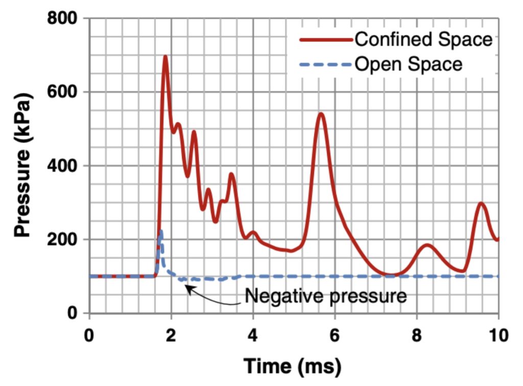
Blast injury happens directly due to the blast wave propagation through the tissue, where the rapid overpressure waves generated by the explosions cause intracranial pressure to increase (Moore et al. 2009; Rezaei et al. 2014). It is this pressure wave that initiates the damage. These waves also induce acceleration, shear stress, skull deformation, and contribute to skull flexure which can lead to brain injury even at lower pressure of 1 bar. The space type is also pertinent, where in confined environments the wave hits a wall, bounces back, and can hit a person again, prolonging exposure (Figure 1). Brain kinematics, on the other hand, describe how the brain and skull move during a blast. Initially, when the wave hits the head, there are large differences in relative velocities between the brain and skull. On the other hand, open spaces allow the velocities to converge to an almost constant value after 15 ms (Rezaei et al. 2014). This means injury risk may be higher in confined spaces. Further, blast injury is systemic: oscillating pressure waves travel through the blood, transmitting kinetic energy from the blast to the brain (Cernak 2010; Moore et al. 2009).
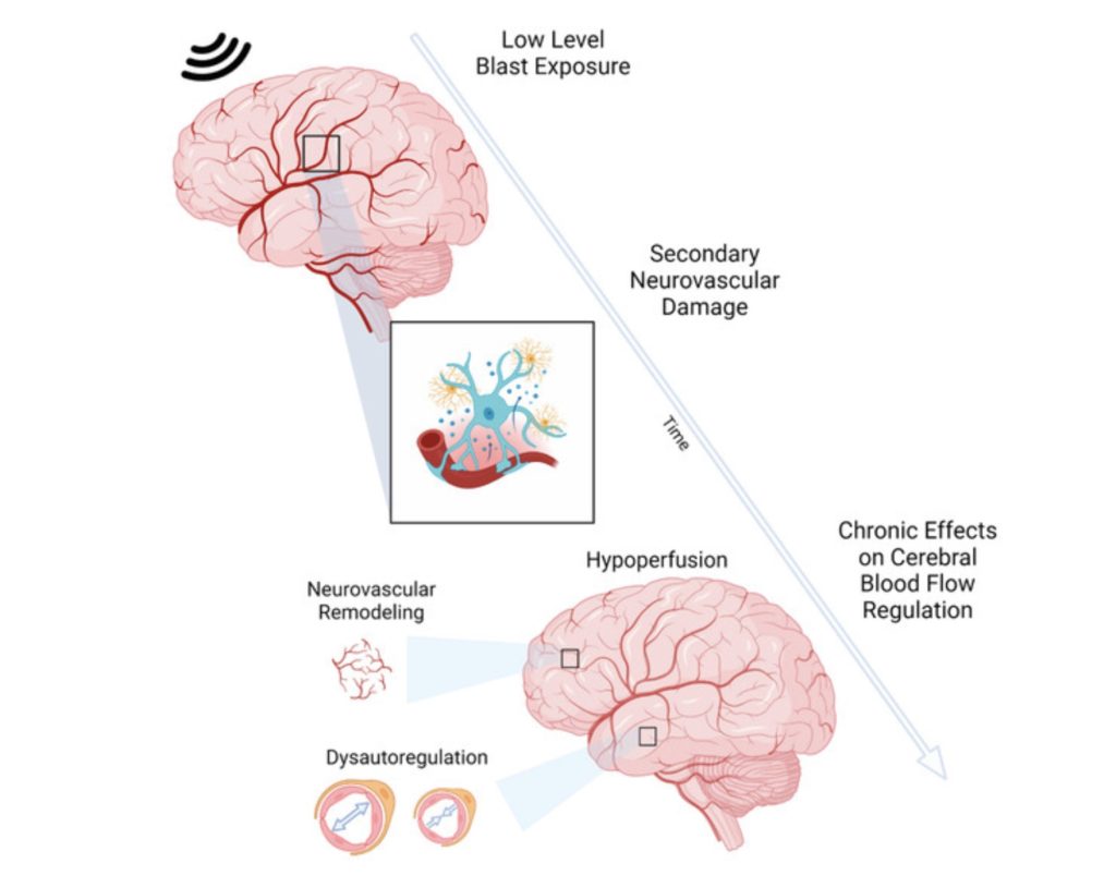
Neurologically, blast neurotrauma is induced by systemic, local, and cerebral responses (Cernak 2010). The way that the injury propagates throughout the brain is through activation of the autonomic nervous system and neuroendocrine–immune pathways. Mechanistically, hyperinflation of the lungs stimulates vagal fibers, triggering a vago-vagal reflex, which can cause widespread cerebral effects and metabolic disruption (Cernak 2010). There can also be neuroinflammation, which may involve axonal injury in white matter regions of the brain. Activated microglia can then contribute to further neural damage through inflammatory signaling (Xu et al. 2016). This inflammation plays an important role in the pathogenesis of long-term neurological deficits. Further, low-level blast exposure, from firing heavy weapons systems or explosives, can contribute to neurological dysfunction (Figure 2) (Kilgore and Hubbard 2024).
Exploring blast-induced neurotrauma is of grave importance because clinical evidence is limited, especially for primary blast exposure (Sachdeva and Ganpule 2024). One such gap in clinical research is in multiple or repetitive blast being more investigated compared to single exposure. That is not to say there is no research. There are findings showing low-level primary blast producing neurological effects like tinnitus, sleep imbalance, cognitive impairment, anxiety, insomnia, and headaches. There is also use of neuroimaging to detect structural changes and loss of white matter integrity (Sachdeva and Ganpule 2024). Nonetheless, more research is needed to minimize existing diagnostic challenges.
Altogether, primary blast injury can be dangerous even for low-level blasts, and the need for progress in diagnosis of brain injury warrants further research into possible detection methods. This can improve care when it comes to military personnel exposed to these blasts in training or combat.
References
Cernak, Ibolja. 2010. “The Importance of Systemic Response in the Pathobiology of Blast-Induced Neurotrauma.” Frontiers in Neurology 1. https://doi.org/10.3389/fneur.2010.00151.
Kilgore, Madison O., and W. Brad Hubbard. 2024. “Effects of Low-Level Blast on Neurovascular Health and Cerebral Blood Flow: Current Findings and Future Opportunities in Neuroimaging.” International Journal of Molecular Sciences 25 (1): 642. https://doi.org/10.3390/ijms25010642.
Moore, David F., Antoine Jérusalem, Michelle Nyein, Ludovic Noels, Michael S. Jaffee, and Raul A. Radovitzky. 2009. “Computational Biology — Modeling of Primary Blast Effects on the Central Nervous System.” NeuroImage 47 (August): T10–20. https://doi.org/10.1016/j.neuroimage.2009.02.019.
Rezaei, A., M. Salimi Jazi, and G. Karami. 2014. “Computational Modeling of Human Head under Blast in Confined and Open Spaces: Primary Blast Injury.” International Journal for Numerical Methods in Biomedical Engineering 30 (1): 69–82. https://doi.org/10.1002/cnm.2590.
Sachdeva, Tarun, and Shailesh G. Ganpule. 2024. “Twenty Years of Blast-Induced Neurotrauma: Current State of Knowledge.” Neurotrauma Reports 5 (1): neur.2024.0001. https://doi.org/10.1089/neur.2024.0001.
Xu, Leyan, Michele L. Schaefer, Raleigh M. Linville, et al. 2016. “Neuroinflammation in Primary Blast Neurotrauma: Time Course and Prevention by Torso Shielding.” Experimental Neurology 277 (March): 268–74. https://doi.org/10.1016/j.expneurol.2016.01.010.