“It was much pleasanter at home,” thought poor Alice, “when one wasn’t always growing larger and smaller, and being ordered about by mice and rabbits.” (Alice in Wonderland, Lewis Carroll)
Sitting at your desk, within seconds, your hands balloon to the size of your couch, your bedroom doorway recedes into an infinite tunnel, and the ticking clock on the wall sounds like it is dragging through molasses. Only, you have not gone down a rabbit hole. You have just been diagnosed with a neurological phenomenon, referred to as Alice in Wonderland Syndrome (AIWS), or Todd’s Syndrome (Figure 1) (Weissenstein et al. 2014).
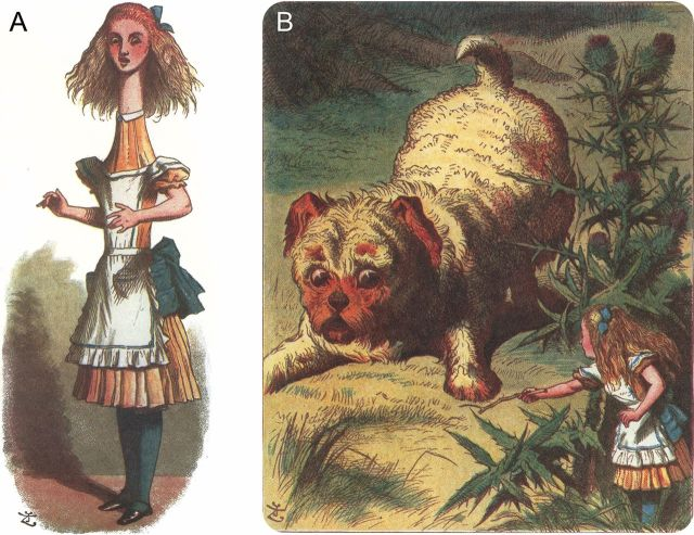
Figure 1: (A) Alice experiences partial macrosomatognosia. (B) Alice experiences total-body microsomatognosia. Illustrations by John Tenniel (Weissenstein et al. 2014).
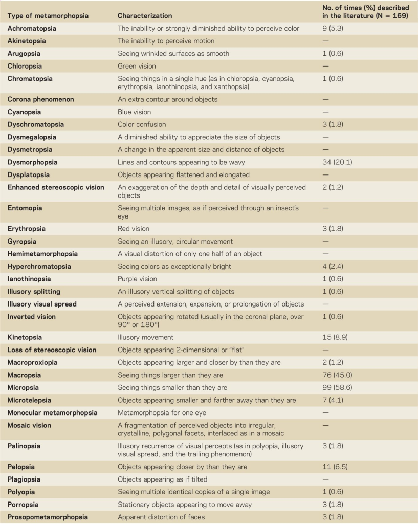
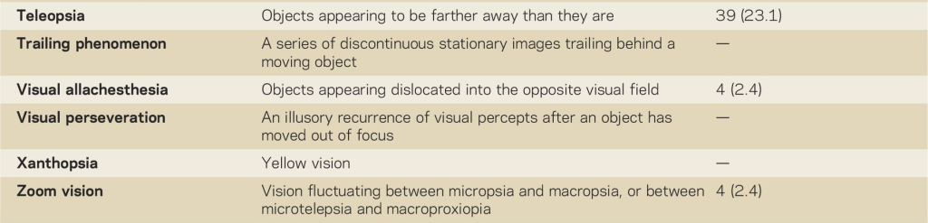
AIWS describes a perceptual disorder characterized by distortions of visual perception (metamorphopsias) of the body schema, external objects, and the experience of time (Blom 2016). AIWS symptoms have expanded to include 42 visual symptoms and 16 somesthetic (or, nonvisual) symptoms (Table 1) (Abbas et al. 2025). Interestingly, these symptoms are characteristic of distortions of sensory perception rather than hallucinations or illusions, rendering the diagnostic and therapeutic consequences of AIWS different from schizophrenia (Blom 2010).
Table 1: Visual distortions that have been described in association with AIWS, with their characteristics and the number of times described in the literature (Blom 2016).
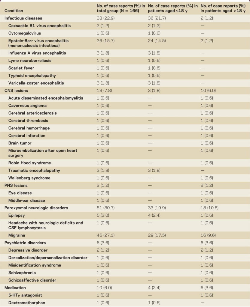
Much of the causes of AIWS is unknown; some sources attribute it to typical migraine, temporal lobe epilepsy, brain tumours, psychoactive drugs, or Epstein-barr-virus infections (Weissenstein et al. 2014). In a systematic review conducted by Blom (2016), the most frequently described condition in which AIWS was associated with was encephalitis, with the Epstein-Barr virus being the most frequently reported pathogen. This was followed by several other neurological conditions, of which migraines were the most frequently reported (Table 2) (Blom 2016).
Table 2: Conditions in which Alice in Wonderland Syndrome has been described in the literature, with their respective number of case descriptions reported by age group (Blom 2016).
To understand what is happening in the brain, researchers have investigated the neuroimagery of several patients reporting AIWS symptoms. In a notable blood-oxygenation level-dependant (BOLD) functional MRI (fMRI) case study of a 12-year-old boy experiencing a viral-onset episode of micropsia (where objects visually shrink), scientists recorded activity directly within the primary and extrastriate visual cortices, alongside increased activation in the parietal lobe (Brumm et al. 2010).
The 12-year old AIWS patient was tasked with several active viewing activities. In one activity, labelled the Ponzo illustration task, participants were asked to make a binary decision as to whether vertical lines were of equal to different lengths. Significantly, the child with AIWS showed more hyperactivation in the right hemisphere superior parietal lobule region of interest (ROI) than the control child (Figure 2). Additionally, the child with AIWS also showed hyperactivation in the inferior parietal cortex ROI relative to the control child. Thus, this study concluded that primary and extrastriate visual cortex regions in the occipital lobe do not respond normally to increased demands for visual analysis during micropsia, particularly during an active viewing task (Brumm et al. 2010).
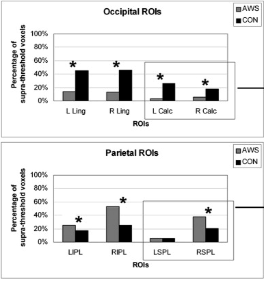
Figure 2: Mean percentage of BOLD signal change for each ROI while viewing the Ponzo Illusion for the child with Alice in Wonderland syndrome (AWS) and the control participant (CON). Percentage of supra-threshold voxels refers to the 3D pixel whose statistical value passes a predefined numerical threshold. Statistically significant differences between participants for each ROI are indicated by an asterisk.
AIWS naturally begs the question: was Lewis Carroll merely an imaginative genius, or was he chronicling his own severe migraine auras? Given his well-documented struggles with debilitating migraines accompanied by visual disturbances (Podoll and Robinson 1999), it seems entirely possible that Carroll didn’t just invent Wonderland – he simply wrote down what his brain was telling him while it was mad as a hatter.
References
Abbas, Yadgar N., Meer M. Abdulkarim, Mohammed Q. Mustafa, et al. 2025. “Unusual Presentation of Alice in Wonderland Syndrome: A Case Report and Literature Review.” Barw Medical Journal 3 (4). https://www.barwmedical.com/index.php/BMJ/article/view/205#references.
Blom, Jan Dirk. 2010. A Dictionary of Hallucinations. Springer.
Blom, Jan Dirk. 2016. “Alice in Wonderland Syndrome: A Systematic Review.” Neurology Clinical Practice 6 (3): 259–70. https://doi.org/10.1212/CPJ.0000000000000251.
Carroll, Lewis, and Donald J. Gray. 2013. Alice in Wonderland. Third edition. A Norton Critical Edition. W. W. Norton & Company.
Podoll, Klaus, and Derek Robinson. 1999. “Lewis Carroll’s Migraine Experiences.” The Lancet 353 (9161): 1366. https://doi.org/10.1016/S0140-6736(05)74368-3.
Weissenstein, Anne, Elisabeth Luchter, and M. A. Stefan Bittmann. 2014. “Alice in Wonderland Syndrome: A Rare Neurological Manifestation with Microscopy in a 6-Year-Old Child.” Journal of Pediatric Neurosciences 9 (3): 303–4. https://doi.org/10.4103/1817-1745.147612.
Comments
10 Responses to “Down the Rabbit Hole We Go: Alice in Wonderland Syndrome”
Hi Leah!
I loved reading your blog post, I have never heard of Alice in Wonderland Syndrome before.
I have a couple suggestions:
P1S2 – I think you are missing a period at the end of the sentence
I noticed that your blog post seems a bit short you could consider expanding on the points in the last paragraph to make it a tad longer.
Might I suggest as well making your references heading larger as well, for me it gets lost a bit in the rest of your writing
Also make sure to add a comment about how it connects to iSci!
Overall an amazing job and I can’t wait to read the final draft!
-Emily
Hi Emily,
Thank you so much for your feedback! I have expanded my blog post a bit more as per your suggestion, and addressed the other feedback. Thank you so much for reading my blog post, I appreciate your edits!
Leah
Hi Leah! This was such an interesting blog post. Below are a few suggestions from me:
-don’t forget to add your comment explaining how your blog post relates to iSci
-I recommend enlarging the references title to better distinguish them from your body text
-If word count permits, you could go into more detail about the physical changes which appear in the brain among someone with AIWS compared to a normal brain
Otherwise, it was a very interesting blog post and you did an amazing job!
Celine
Hi Celine,
Thank you so much for your suggestions! I have tried my best to implement them all. Hopefully, it’s a little bit more clear now regarding the difference between normal vs AIWS brain!
Thanks for reading my blog post!
Leah
Hi Leah!
I really enjoyed reading this! I had never heard of Alice in Wonderland Syndrome before, and I loved how you used the Alice in Wonderland imagery throughout the post. It made such a strange neurological phenomenon really easy to picture 🙂
I had a few suggestions while I was reading:
– In your third paragraph, you say that the fMRI case study “proved” that the distortions come from transient disruptions in how the brain processes space and scale. Since this was a case study of one 12-year-old patient, I wonder if “provided evidence” or “suggested” might be a little more accurate than “proved.” I think that would make the wording better reflect what can be concluded from the study.
– I really like the Alice in Wonderland theme, but I noticed you use “mad” both in the opening and again in your final sentence. Since you later discuss the psychological experiences of people with AIWS, I wonder if that wording could unintentionally come across as a little stigmatizing. You could definitely keep the humour while rewording those parts slightly, especially the final line! Completely up to you though 🙂
– For Figure 2, the caption refers to “Alice in Wonderland syndrome (AWS),” while you use AIWS everywhere else in the post. I would keep the abbreviation consistent or briefly explain if AWS is the wording used in the original study. It might also help readers understand the figure if you briefly point out what difference between the AIWS participant and control they should actually be looking for.
Overall, this was such an interesting read and I really liked the way you connected the neurological and psychological sides of AIWS!
Happy editing!
Saniyah
Hi Saniyah,
Thank you so much for your detailed feedback! This was very helpful! I have tried my best to implement your suggestions. I hope my use of the term “mad” didn’t come across as stigmatizing; I only meant it as a play on Lewis Carroll’s “mad hatter.” I will definitely check this over, though, and I really appreciate you pointing this out!
Thank you again for reading my post and for your detailed feedback!
Leah
Hi Leah! I enjoyed reading your blog post, I found it very well written, it had a lovely structure and flow.
Here are some minor fixes to consider:
– Lewis Carroll should be spelt with 2 Ls (after the quote, figure description, last sentence)
– P3S3 unless you are certain, I would avoid saying ‘proved.’ – if you are not certain, you could instead say: ‘supported the hypothesis that’ ‘suggested that’ ‘provided evidence that’ etc.
– Also like others said, don’t forget to make your comment connecting your post to iSci
Happy editing!
– Hafsah S
Hi Hafsah,
Thank you so much for your detailed feedback on my blog post! I hope I have done my best to ensure I have implemented all your suggestions!
Thanks again for reading my blog post!
Leah
Hi Leah,
This was a really interesting read. I especially liked your title, it caught my attention immediately. I just have a few minor suggestions:
– I would consider explaining what these terms mean “metamorphopsias (such as macropsia and micropsia), somatognosic disturbances, and chronometric alterations”
– You mention that AIWS “manifests as a severe variant of a migraine aura.” I was wondering whether AIWS is considered a specific type of migraine aura, or whether it is more accurately described as a syndrome that can be associated with migraine. It may be helpful to adjust the wording to clarify this distinction.
Other than that, I really enjoyed your post and I look forward to reading the final version!
Ananya
Hey iSci,
I decided to write this blog post because not only have I recently started reading Lewis Carroll’s original, “Alice in Wonderland” novel, but this topic came up in one of my elective courses. Not much is known to this day about Alice in Wonderland syndrome, and so I wanted to briefly investigate associated symptoms and neurological conditions. I also found that this topic uniquely related to some first-year psychology and second-year neuroscience content in iSci!
Please let me know your feedback and suggestions; I’m looking forward to your advice for edits!
Leah