The human brain contains billions of neurons, intricate networks of nerve fibres, and structures hidden beneath a hard skull. Yet doctors can view all these, without making a single incision, using… magnets? Magnetic resonance imaging (MRI) makes this possible by detecting signals from hydrogen nuclei within the body. Rather than directly photographing the brain, MRI uses magnetic fields to produce signals that computers then transform into detailed images (Plewes and Kucharczyk 2012). But how can a magnet create an image of something hidden inside our heads?
It starts with hydrogen, which is abundant in our body’s water and fat. Each hydrogen nucleus contains a proton with a property called spin, making it act like a tiny magnet. Normally, these magnets point in different directions. Inside an MRI scanner, however, slightly more protons align with the scanner’s magnetic field than against it. This creates a small amount of net magnetization (Azhar and Chong 2023).
Subsequently, the scanner sends radio frequency pulses into the body, transferring energy to the hydrogen nuclei and tilting their net magnetization. When the pulses stop, the nuclei gradually return to equilibrium, producing changing magnetic signals detected by the scanner’s receiving coils (Plewes and Kucharczyk 2012).
Here’s where it gets interesting though, If hydrogen is everywhere in the brain, why doesn’t an MRI show one big grey image? The answer lies in the different environments surrounding hydrogen nuclei. Two processes explain this; T1 relaxation, which describes how quickly magnetization recovers along the main magnetic field, while T2 relaxation describes how quickly the nuclei lose synchronization with one another. Both depend on interactions with nearby molecules (Gaeta et al. 2024).
For example, hydrogen in freely moving water behaves differently from hydrogen in water interacting with proteins and fats. These surroundings affect how quickly signals recover and fade, so different tissues produce different MRI signals (Gaeta et al. 2024).
This is how MRI distinguishes brain structures. White matter contains fatty myelin around nerve fibres, while grey matter contains more neuronal cell bodies. Cerebrospinal fluid is mostly water. Because their compositions differ, so do their relaxation times. In T1-weighted images, white matter usually appears brighter than grey matter, while cerebrospinal fluid looks dark. In T2-weighted images, cerebrospinal fluid appears bright (Azhar and Chong 2023).
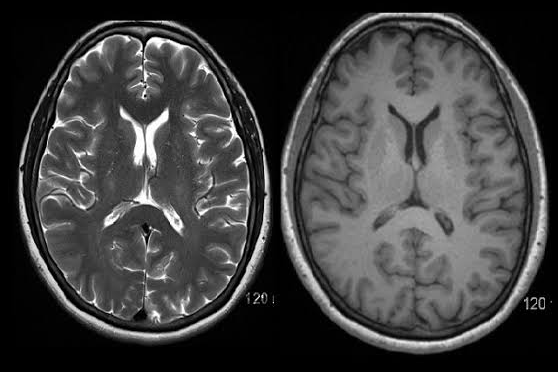
Figure 1. Comparison of T1-weighted (right) and T2-weighted (left) MRI scans of the human brain, showing differences in tissue brightness caused by varying relaxation times.
How does the scanner know where each signal comes from? It uses magnetic-field gradients, which slightly change the magnetic field across different locations. As a result, signals from different parts of the brain have different frequencies and phases. A computer uses this information to reconstruct an image (Plewes and Kucharczyk 2012).
Doctors can also adjust the timing of radiofrequency pulses and signal collection to highlight different tissue properties. Comparing these MRI images helps them identify abnormalities, including possible tumours that look different from healthy tissue (Azhar and Chong 2023).
The next time you see an MRI scan, remember that it isn’t simply a photograph of the brain. It is an extraordinary image created by transforming the invisible magnetic behaviour of atomic nuclei into something the human eye could never see on its own.
References
Azhar, Syifa, and Le Roy Chong. 2023. “Clinician’s Guide to the Basic Principles of MRI.” Postgraduate Medical Journal 99 (1174): 894–903. https://doi.org/10.1136/pmj-2022-141998.
Gaeta, Michele, Karol Galletta, Marco Cavallaro, Enricomaria Mormina, Maria Teresa Cannizzaro, Ludovica Rosa Maria Lanzafame, Tommaso D’Angelo, Alfredo Blandino, Sergio Lucio Vinci, and Francesca Granata. 2024. “T1 Relaxation: Chemo-Physical Fundamentals of Magnetic Resonance Imaging and Clinical Applications.” Insights into Imaging 15 (1): Article 200. https://doi.org/10.1186/s13244-024-01744-2.
Plewes, Donald B., and Walter Kucharczyk. 2012. “Physics of MRI: A Primer.” Journal of Magnetic Resonance Imaging 35 (5): 1038–54. https://doi.org/10.1002/jmri.23642.


Leave a Reply
You must be logged in to post a comment.