If you’ve ever experienced an ultrasound imaging procedure, it can seem a bit silly. In a dark, clinical room, cold gel is put on an area of your body as a radiologist or sonographer solemnly probes at you with a transducer, the component of the ultrasound machine that converts electricity into ultrasound (Mayo Clinic 2024; Suetens 2017, 148). But what exactly is ultrasound, and what properties allow it to be used in medical imaging and beyond?
Firstly, sound energy travels in longitudinal waves of compressional and rarefactional air pressure deviations from atmospheric temperature, such as those created from banging a drum and moving the air around it (Photinos 2021, 20). The frequency of sound waves mainly depends on the density of the medium of propagation. When sound waves reach a density difference between two mediums, diffuse reflection – or unfocused dispersion – of the waves can occur (Photinos 2021, 34-37).
Ultrasound waves are considered to be sound waves over 20,000 Hz or cycles a second – above the range of sound detection in humans (Porteous 2008, 595). Ultrasound can be generated through the inverse piezoelectric effect, where an applied voltage leads to the deformation of a crystal such as lead zirconate titanate or polyvinylidene fluoride, generating waves of air compression and rarefaction at high frequencies (Suetens 2017, 148-156). These high frequency waves and their ability to reflect when impeded by a medium boundary allow for the precise measurement used in medical imaging to create a sharp image.
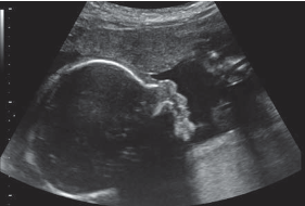
The three-dimensional image of an ultrasound scan is generated when the mediums the sound waves travel through are mapped by analyzing the reflection at boundaries of tissue. This differentiation of tissue is determined by the Doppler effect and its effect on the reflected waves (Suetens 2017, 147-154).

The Doppler effect (Figure 2.) used in tandem with measuring distance using the time taken for the reflected wave to be detected, the raw data can be computationally mapped into a three-dimensional image (Figure 1.) that can be used for monitoring pregnancy and evaluating bone and joint health (Suetens 2017, 147-183; Mayo Clinic 2024).
But ultrasound has additional critical uses beyond imaging and radiology. One notable example of this is therapeutic ultrasound. High-intensity focused ultrasound (HIFU) can be used to ablate or destroy benign tumors by rapidly generating heat at a small section of the tissue to induce necrosis or death of the targeted tissue (Cheung, 2018). An expansion of HIFU beyond targeting soft tissue is histotripsy, a procedure used to treat kidney stones by eroding them into small pieces that are more easily passed (Xu 2024).
Ultrasound and the physics behind it prove critical in modern medical imaging and treatments. The same sound technology that can be used to find a growth on the inside of your body can also ablate the very same growth – neat! The research is sound: ultrasound is ultra-useful in medicine today.
References
Cheung, Vincent Y. T. “High-Intensity Focused Ultrasound Therapy.” Best Practice & Research. Clinical Obstetrics & Gynaecology (Netherlands) 46 (January 2018): 74–83. https://doi.org/10.1016/j.bpobgyn.2017.09.002.
Mayo Clinic. 2024 “Ultrasound.” Mayo Foundation for Medical Education and Research. May 3, 2024. Accessed October 28th, 2026. https://www.mayoclinic.org/tests-procedures/ultrasound/about/pac-20395177.
Photinos, Panos, and publisher Institute of Physics. The Physics of Sound Waves : Music, Instruments, and Sound Equipment. Second edition. IOP Publishing, 2021. https://doi.org/10.1088/978-0-7503-3539-3.
Porteous, Andrew. Dictionary of Environmental Science and Technology, John Wiley & Sons, Incorporated, 2008. ProQuest Ebook Central, https://ebookcentral.proquest.com/lib/mcmu/detail.action?docID=470320.
Suetens, Paul. “Ultrasound Imaging.” Chapter. In Fundamentals of Medical Imaging. Cambridge University Press, 2017.
Xu, Zhen, Tatiana D. Khokhlova, Clifford S. Cho, and Vera A. Khokhlova. “Histotripsy: A Method for Mechanical Tissue Ablation with Ultrasound.” Annual Review of Biomedical Engineering (United States) 26, no. 1 (2024): 141–67. https://doi.org/10.1146/annurev-bioeng-073123-022334.


Leave a Reply
You must be logged in to post a comment.