In archaeology, material characterization is often fundamental to identification and appraisal of artifacts. For example, authenticity assessments of Chinese jade requires confirmation of both mineral composition and microstructure to ensure the correct makeup of tremolite and actinolite (Zhang et al. 2020). However, commonly-used analytical techniques, such as chromatography, require destruction of the sample, which is undesirable for these culturally valuable objects (Musílek et al. 2012). To avoid this, X-ray fluorescence (XRF) and X-ray crystallography can be used; these are non-destructive techniques that use the compound’s “reaction” to X-ray illumination to determine the chemical and structural characteristics of a molecule (Loubser and Verryn 2008).
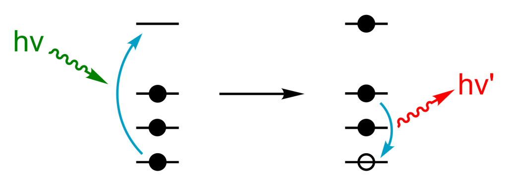
XRF is an analytical technique used to determine chemical composition (Marguí et al. 2022). X-ray exposure can cause atoms to ionize, assuming the energy of the X-ray is greater than the ionizing energy of the atom. This ionization causes the atom to eject an electron, leaving behind an orbital hole. To fill this space, an electron in a higher orbital will “fall” down into the hole, causing a release of energy (as a photon) that is equal to that energy difference (Figure 1). The energy of this radiation is characteristic for each element, meaning the resultant signals can be interpreted to determine the elemental composition. The intensity of the radiation can further be used to determine the relative amount of each element within the sample. However, this analysis does not give us the full picture; XRF tells us composition, but the compound of interest could be any of several different isomers (Marguí et al. 2022).
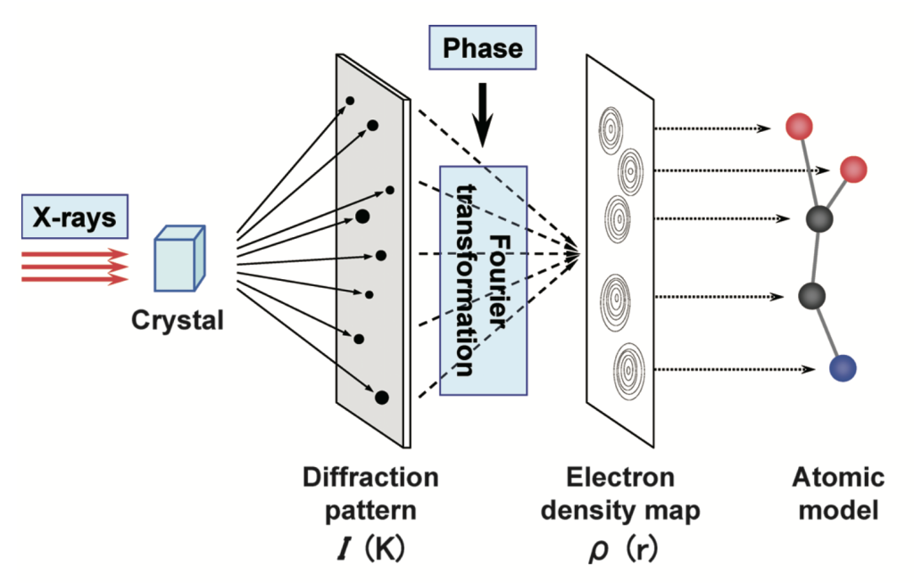
To gain a more detailed understanding of the compound, X-ray crystallography is used. This technique uses X-ray diffraction (XRD) and thus requires a crystal sample (Ameh 2019). This time, the X-rays are diffracted by the sample, producing a two-dimensional diffraction pattern. The crystal is slowly rotated, with pattern recordings taken at each orientation. The position and intensities of the spots in these patterns directly correlate to the dimensions of the unit cell, which is the smallest repeating unit within the crystal. The amplitude and phase of the scattered X-rays can further be determined from the diffraction pattern. A Fourier transform can then convert these two-dimensional patterns to a three-dimensional electron density map. This map defines the electron density at every point within the unit cell, with the maxima taken as the atoms in the sample. This creates the preliminary model of the structure, which is then refined using complementary chemical information (including the chemical formula, from XRF) to produce the final crystal structure (Figure 2; Ameh 2019).
Accurate techniques still possess limitations. For example, in practice, elements lighter than sodium often cannot be characterized by XRF (Marguí et al. 2022). Further, XRD requires a single-crystal sample, meaning it has a continuous, unbroken lattice, which is not possible for all compounds (Ameh 2019). Consequently, XRF cannot be used to identify organics, but is key to the composition analysis of ceramics and metals to identify possible trade routes (Musílek et al. 2012). Similarly, XRD cannot be used for non-crystalline materials, but provides a detailed analysis of mineral inclusions or corrosion products in artifacts, which can be used for origin determination and dating, respectively (Garrison 2014). Regardless, both techniques are incredibly valuable in material characterization and continue to be the gold standard in archeological applications.
References
Ameh, E. S. 2019. “A Review of Basic Crystallography and X-Ray Diffraction Applications.” The International Journal of Advanced Manufacturing Technology 105 (7–8): 3289–302. https://doi.org/10.1007/s00170-019-04508-1.
Calvero. 2006. Schematic Representation of X-Ray Fluorescence (XRF). Selfmade with ChemDraw. https://commons.wikimedia.org/wiki/File:X-ray_fluorescence_simple_figure.svg.

Garrison, Ervan. 2014. “X-Ray Diffraction (XRD): Applications in Archaeology.” In Encyclopedia of Global Archaeology. Springer, New York, NY. https://doi.org/10.1007/978-1-4419-0465-2_339.
Hasegawa, Kimiko. 2012. “Introduction to Single Crystal X-Ray Analysis.” The Rigaku Journal 28 (1): 5.
Kuckova, Stepanka, Ivan Nemec, Radovan Hynek, Janka Hradilová, and Tomas Matys Grygar. 2005. “Analysis of Organic Colouring and Binding Components in Colour Layer of Art Works.” Analytical and Bioanalytical Chemistry 382 (June): 275–82. https://doi.org/10.1007/s00216-005-3108-5.
Loubser, Maggi, and Sabine Verryn. 2008. “Combining XRF and XRD Analyses and Sample Preparation to Solve Mineralogical Problems.” South African Journal of Geology – S AFR J GEOL 111 (September): 229–38. https://doi.org/10.2113/gssajg.111.2-3.229.
Marguí, E., I. Queralt, and E. de Almeida. 2022. “X-Ray Fluorescence Spectrometry for Environmental Analysis: Basic Principles, Instrumentation, Applications and Recent Trends.” Chemosphere 303 (September): 135006. https://doi.org/10.1016/j.chemosphere.2022.135006.
Musílek, Ladislav, Tomáš Cechák, and Tomáš Trojek. 2012. “X-Ray Fluorescence in Investigations of Cultural Relics and Archaeological Finds.” Applied Radiation and Isotopes: Including Data, Instrumentation and Methods for Use in Agriculture, Industry and Medicine 70 (7): 1193–202. https://doi.org/10.1016/j.apradiso.2011.10.014.
Zhang, Baoshuai, Xiaotong Wu, Yufeng Sun, et al. 2020. “Complex Raw Materials and the Supply System: Mineralogical and Geochemical Study of the Jade Artefacts of the Longshan Culture (2400–2000 Bce) from Sujiacun Site in Coastal Shandong, China.” Archaeometry 63 (December): 1–18. https://doi.org/10.1111/arcm.12634.
Comments
9 Responses to “What’s Inside? Non-Destructive Analysis with XRD and XRF”
Hi iSci! My group’s Terroir article for WineSci investigated irradiative techniques for wine preservation, which included X-ray irradiation. This got me thinking about other applications of X-rays, and led me to some pretty cool topics at the centre of chemistry and physics!
– Amelie
Hi Amelie,
This blog was very well-written. Your explanations about XRF and XRD were very clear. Here are a few suggestions:
1. First paragraph, first sentence: You could provide examples of a few fields or highlight a famous object that was characterized and damaged in the process. This would create a more captivating beginning!
2. Last paragraph, third sentence: You describe a single-crystal sample in brackets, but it might improve flow if you directly included the defintion. You could say “Further, XRD is not always feasible as it requires a single-crystal sample, meaning the sample has a continuous, unbroken lattice.”
3. Last paragraph, second last sentence: What is provenance determination? If word count allows, maybe you could include a bit about this.
Good luck editing,
Ria
Hi Ria! Thanks for your comment — I have edited my intro to include a tidbit about Chinese jade appraisal, and removed the brackets per your recommendation. Also, I’ve changed the word provenance to origin, as it is more commonly known. Thanks 🙂
Hello Amelie,
This was a very interesting read! You managed to clearly explain how artifacts can be analyzed without physical touch, while bringing in concepts from all the way from first-year.
A few suggestions:
1) While your explanation of the XRF and crystallography were very well done, if you can find a way to give a recent example of when these techniques may have been used to identify something interesting about an artifact, that would make this article even more engaging.
2) You state that it is “not always realistic” for the use of a single-crystal sample, but it’s hard to understand that reasoning. If it’s in those brackets nearby, I would recommend adjusting the wording to allow for a better flow. If there are other reasons, it would be best if you explicitly state the reasons, briefly.
3) P2S4: Probably just an aesthetic preference for myself, but two sets of parentheses beside each other looks awkward. Perhaps you could find a way to remove the parentheses before the citation by adding a few words to the end of the sentence.
I know I’m being nitpicky, so let me reiterate that this was a superb article!
– Liam
Hi Liam! Thanks for your comment! I have added a tidbit to my intro about authentication of Chinese jade artifacts, and clarified that single-crystal samples do not exist for all compound types. I also deleted the add-on in brackets to avoid the double brackets in P2S4. Thanks!
Hi Amelie, great blog post. This was a new topic for me so I found it very interesting! Here are a few things I would suggest to help improve your post.
In the P1S1, i think it needs a reword, or it needs something added to it, it just feels a little unfinished. Maybe you can add some examples?
You may want to remove the hyperlinks in your references to make it look more professional.
Besides that i think this was an amazing blog post! Happy editing
Brianna
Hi Brianna! Thanks for your feedback! The SciLit component outlines indicates that hyperlinked DOIs are preferred, which is why I made that choice. I also rewrote the intro to include some examples. Thanks!
Really great blog post! As someone currently struggling through orgo, I appreciate learning something a little more interesting about organic chemistry. I just had a few minor suggestions to improve your post:
– Make sure to centre your figure captions.
– In Chicago style, it should be (Fig. 1) instead of (Figure 1)
– In your second reference, the year and month are separated, so I suggest double-checking that your citation generator spit that out properly, as Chicago usually doesn’t even include the month at all.
Happy editing!
Hi Sarah! Thanks for your comments 🙂 I have centred my figure captions and fixed that citation, thanks for catching that! Also, after reviewing the manual, it seems that the Fig. vs Figure choice is preference, and so I’m going to stick with spelling it out 🙂 Thanks!