Have you ever wondered how your brain works, how thoughts turn into actions, and how new ideas arise? This incredible process can be explained by analyzing the electrical and chemical processes which permit neuronal communication.
If we consider the general steps a neuron goes through, we can observe that they behave like biological batteries. At rest, the strong electrical potential, about -70 mV, is caused by an uneven distribution of ions across the lipid bilayer (Wright 2004). In particular, the negative voltage is due to the fact that the membrane is selectively permeable, and allows K+ to leak out, leaving behind a negative charge. At rest, sodium-potassium pumps use ATP to maintain this potential, pushing Na+ out and K+ in, against their concentration gradients (Skou 1998). This process is crucial, as it maintains the conditions necessary for neurons to quickly fire when signals are received.
Beyond this, neurons can be thought of as parallel resistor-capacitor (RC) circuits, where the ion channels act as the resistors, while the lipid bilayer acts as the capacitor (Dabrowski et al. 2013). More specifically, the protein channel in the membrane is a variable resistor, which controls an ion’s ability to flow across according to Ohm’s Law:
I = V/R
Equation 1. Ohm’s Law equation used in circuits, where I is the current of electrical charge in amperes, V is the voltage, or electrical pressure, in volts, and R is the resistance in ohms.
By examining Equation 1. we can see that when voltage is constant, current and resistance are inversely proportional. As a result, when the channel is open, allowing ions to flow, the resistance decreases, while the current increases, amplifying the conductivity (Dabrowski et al. 2013). Moreover, the lipid membrane is a leaky capacitor, which acts like a non conductive insulator, separating positively and negatively charged ions on opposing sides, like a capacitor plate (El-Beyrouthy and Freeman 2021). However, this barrier is leaky, meaning that some charge will pass through, but this amount is insignificant, and quickly fades. Since this electric signal is too weak, and dwindles, the axon requires some sort of spike when it actually wants to fire. This is where our action potential comes in.
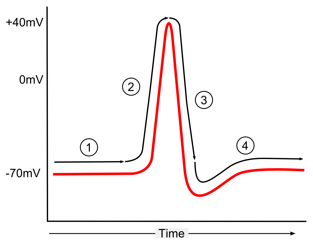
Figure 1. Action potential graph showing the changes in a neuron’s potential energy (mV) as time passes. The graph emphasizes the different phases, as the cell begins at resting potential (1), depolarizes to the max peak (2), repolarizes back down (3), briefly hyperpolarizes, and finally returns to the original state (4) (Duncan 2017).
As seen in Figure 1, when the voltage reaches the threshold, about -55 mV, the neuron will fire, following the all-or-nothing principle. This is due to the fact that S4 helix, a voltage sensor, detects that the threshold has been met, and opens the ion channels, lowering the membrane resistance, and allowing Na+ to rush in (Catterall 2010; Bean 2007). This causes the spike seen in Figure 1, due to a change in the electrical charge distribution, leaving the voltage at around +40 mV. Once the peak is achieved, sodium gates are no longer active, preventing ions from flowing across the membrane, while potassium channels, including Kv3 and Kv4, allow K+ to leave the neuron, briefly hyperpolarizing the neuron (Bean 2007). The sodium-potassium pumps will then move K+ into the cell, and Na+ out, restoring the resting potential.
How does this all connect to how your brain works? The firing of one neuron completes the circuit, and leads to the firing of another, and so on, allowing for motion, thinking, and more. Overall, these small-scale reactions take place all over the body, spreading millivolts throughout, providing insight to human consciousness. Ultimately, our thoughts and movements are, as far as we know, a series of electrical impulses propagating throughout our bodies.
References
Bean, Bruce P. 2007. “The Action Potential in Mammalian Central Neurons.” Nature Reviews Neuroscience 8 (6): 451–65. https://doi.org/10.1038/nrn2148.
Catterall, William A. 2010. “Ion Channel Voltage Sensors: Structure, Function, and Pathophysiology.” Neuron 67 (6): 915–28. https://doi.org/10.1016/j.neuron.2010.08.021.
Dabrowski, Katie M, Diego J Castaño, and Jaime L Tartar. 2013. “Basic Neuron Model Electrical Equivalent Circuit: An Undergraduate Laboratory Exercise.” Journal of Undergraduate Neuroscience Education 12 (1): A49–52. https://pmc.ncbi.nlm.nih.gov/articles/PMC3852871/.
Duncan, Peter. 2017. Action Potential Schematic Showing 1) Resting Membrane Potential 2) Depolarisation 3) Repolarisation and 4) Hyperpolarisation and Return to Baseline. In Wikimedia Commons. June 1. https://upload.wikimedia.org/wikipedia/commons/e/e0/Action_potential_schematic.png?utm_source=commons.wikimedia.org&utm_campaign=index&utm_content=original.
El-Beyrouthy, Joyce, and Eric Freeman. 2021. “Characterizing the Structure and Interactions of Model Lipid Membranes Using Electrophysiology.” Membranes (Switzerland) 11 (5): 319. PubMed Central. https://doi.org/10.3390/membranes11050319.
Skou, Jens Christian. 1998. “The Identification of the Sodium Pump.” Bioscience Reports 18 (4). https://web.as.uky.edu/Biology/faculty/cooper/ABLE-2021/ABLE-2021-MembranePotenial-tempAndK/Nobel%20prize%20Na-K%20pump.pdf.
Wright, Stephen H. 2004. “Generation of Resting Membrane Potential.” Advances in Physiology Education 28 (4): 139–42. https://doi.org/10.1152/advan.00029.2004.


Leave a Reply
You must be logged in to post a comment.