Over the past several decades, advances in neuroimaging have revealed that the human brain is not a uniform processing system. One particularly intriguing example is face recognition.
Despite how effortless it feels, identifying a face requires the brain to rapidly distinguish subtle visual features from a vast amount of sensory information. This has led neuroscientists to investigate whether the brain contains dedicated neural circuitry for face perception.
Research suggests that face perception is not simply another form of object recognition. Instead, humans appear to possess specialized neural mechanisms dedicated to processing faces. Central to this system is the fusiform face area (FFA), a region of the temporal lobe that plays a crucial role in identifying and interpreting faces (Kanwisher, 2006)
Faces are among the most important visual stimuli humans encounter. Within fractions of a second, we can determine a person’s identity, emotional state, approximate age, sex, and even where they are looking.
Because facial recognition is so important, neuroscientists have long debated whether the brain contains specialized “face-processing” regions or whether general visual mechanisms are sufficient. This debate is known as the face-specificity hypothesis (Kanwisher, 2006).
Rather than responding to visual complexity alone, the FFA appears specialized for detecting and recognizing faces, as demonstrated by phenomena such as the face inversion effect, where upside-down faces are much harder to recognize than inverted objects.
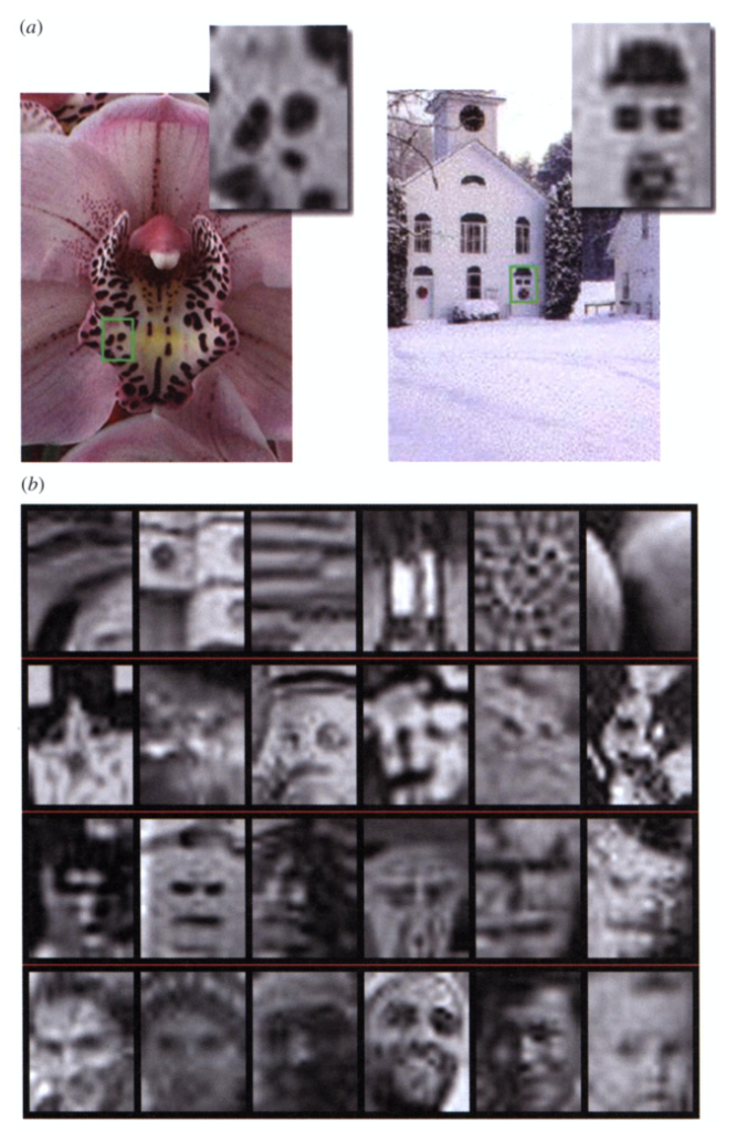
Figure 1. Examples of Stimuli with Varying Degrees of Face Resemblanc. Stimuli ranged from non-face images to genuine faces and were used to investigate left and right fusiform gyrus responses to facial similarity (Meng et al., 2012).
Further support comes from research examining the left and right fusiform gyri. The fusiform gyri are two brain structures located on the underside of the temporal lobes, one in each hemisphere.
Using images that ranged from non-face patterns to genuine faces (Figure 1), researchers found that the right fusiform gyrus categorized stimuli as faces or non-faces, whereas the left fusiform gyrus responded more gradually as facial resemblance increased. These findings suggest that the right hemisphere is specialized for face detection, while the left hemisphere is more sensitive to varying degrees of face-like features (Meng et al., 2012).
The fusiform face area serves as a key component of this network, allowing humans to rapidly detect and recognize faces. By studying how the FFA works, neuroscientists continue to uncover how the brain transforms simple visual input into one of our most socially important abilities: recognizing the people around us
References.
Kanwisher, Nancy, and Galit Yovel. “The Fusiform Face Area: A Cortical Region Specialized for the Perception of Faces.” Philosophical Transactions: Biological Sciences 361, no. 1476 (2006): 2109–28. http://www.jstor.org/stable/20209804.
Meng, Ming, Tharian Cherian, Gaurav Singal, and Pawan Sinha. “Lateralization of Face Processing in the Human Brain.” Proceedings: Biological Sciences 279, no. 1735 (2012): 2052–61. http://www.jstor.org/stable/41549504.
Murty, N. Apurva Ratan, Santani Teng, David Beeler, Anna Mynick, Aude Oliva, and Nancy Kanwisher. “Visual Experience Is Not Necessary for the Development of Face-Selectivity in the Lateral Fusiform Gyrus.” Proceedings of the National Academy of Sciences of the United States of America 117, no. 37 (2020): 23011–20. https://www.jstor.org/stable/26969232.
Ramon, Meike, Luca Vizioli, Joan Liu-Shuang, and Bruno Rossion. “Neural Microgenesis of Personally Familiar Face Recognition.” Proceedings of the National Academy of Sciences of the United States of America 112, no. 35 (2015): E4835–44. https://www.jstor.org/stable/26464134.


Leave a Reply
You must be logged in to post a comment.